(Cat. No. 401502) Human BCMA / TNFRSF17 Protein, Fc Tag, Alexa Fluor 647, 100 tests
Beschreibung
Description
Tumor necrosis factor receptor superfamily, member 17 (TNFRSF17), also known as B-cell maturation antigen (BCMA) or CD269 antigen, is a member of the TNF-receptor superfamily.
This receptor is preferentially expressed in mature B-lymphocytes, and may be important for B-cell development and autoimmune response and has been shown to specifically bind to the tumor necrosis factor (ligand) superfamily, member 13b (TNFSF13BBAFF), and to lead to NF-kappaB and MAPK8/JNK activation.
TNFRSF17/BCMA is a target of donor B-cell immunity in patients with myeloma who respond to DLI. Antibody responses to cell-surface BCMA may contribute directly to tumor rejection in vivo.
Specification
- Vol. per test: 1 µl
- Gene name synonym: BCMA; CD269; TNFRSF17
- Source: Human
- Expression host: ExpiCHO cells
- Molecular mass: The recombinant human TNFRSF17 consists of 303 amino acids and predicts a molecular mass of 33.3 kDa. In SDS-PAGE under reducing conditions, the apparent molecular mass of human TNFRSF17 is approximately 33-44 kDa due to the glycosylation
- Endotoxin: < 1.0 EU per μg protein as determined by the LAL method
- Reactivity: Mouse
- Purity: > 95 % as determined by SDS-PAGE
- Storage buffer:
Aqueous buffered solution containing protein stabilizer and ≤0.05% ProClin 300
Preparation and Storage
- Shipped at 2–8 °C.
- Store undiluted at 2–8 °C and protected from prolonged exposure to light
- Avoid freeze /thaw
- The monoclonal antibody was purified by Protein A
- The antibody was conjugated with Alexa Fluor 647 under optimum conditions and unincorporated dye was removed
Product Notices
- Since applications vary, each investigator should titrate the reagent to obtain optimal results.
- Caution: Reagent containing ProClin 300 should be handled with care. Do not take internally and avoid all contact with the skin, mucosa and eyes.
- This product is provided under an intellectual property license from Life Technologies Corporation. The transfer of this product is conditioned on the buyer using the purchased product solely in research conducted by the buyer and the buyer must not (1) use this product or its components for (a) diagnostic, therapeutic or prophylactic purposes; or (b) manufacturing or quality assurance or quality control, and/or (2) sell or transfer this product or its components for resale, whether or not resold for use in research. For information on purchasing a license to this product for purposes other than as described above, contact Life Technologies Corporation, 5781 Van Allen Way, Carlsbad, CA 92008 USA or outlicensing@thermofisher.com.
- Conditions: The information disclosed herein is not to be construed as a recommendation to use the above product in violation of any patents. Bioswan will not be held responsible for patent infrigement or other violations that may occur with the use of our products. Purchase does not include or carry any right to resell or transfer this product either as a stand-alone product or as a component of another product. Any use of this product other than the permitted use without the express written authorization of Bioswan Company is stickly prohibited.
- For Research Use Only.
- Not for use in diagnostic or therapeutic procedures.
- Not for resale.
- Bioswan, the Bioswan Logo and all other trademarks are property of BioSwan Laboratories, Co., Ltd..
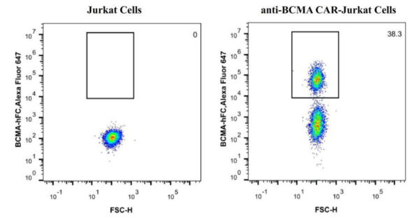
FCM Protocol
- Harvest the cells and wash the cells once by FACS buffer (PBS containing 2% of BSA).
- Count the cells number and the viability, aliquot up to 2×10⁵ live cells into each tube. (Note: the cell viability must be ≥ 95%.)
- Resuspend cells in 100 μL of diluted Human BCMA Protein, Fc Tag, Alexa Fluor 647 (Cat. No. 401501, 1:100 diluted in FACS buffer) for 30 min at 4°C.
- Wash the cells 3 times by FACS buffer and resuspend the cells in 200 μL PBS per sample.
- Transfer the cells into flow tube and analyze on Flow Cytometer. Acquisition of >10, 000 events is performed.